COVID-19 causes unexpected cellular response in the lungs, research finds
New insights into the immune response to SARS-CoV-2 infections could bring better treatments for COVID-19 cases.
An international team of researchers unexpectedly found that a biochemical pathway, known as the immune complement system, is triggered in lung cells by the virus, which might explain why the disease is so difficult to treat. The research is published this week in the journal Science Immunology.
The researchers propose that the pairing of antiviral drugs with drugs that inhibit this process may be more effective. Using an in vitro model using human lung cells, they found that the antiviral drug Remdesivir, in combination with the drug Ruxolitinib, inhibited this complement response.
This is despite recent evidence that trials of using Ruxolitinib alone to treat COVID-19 have not been promising.
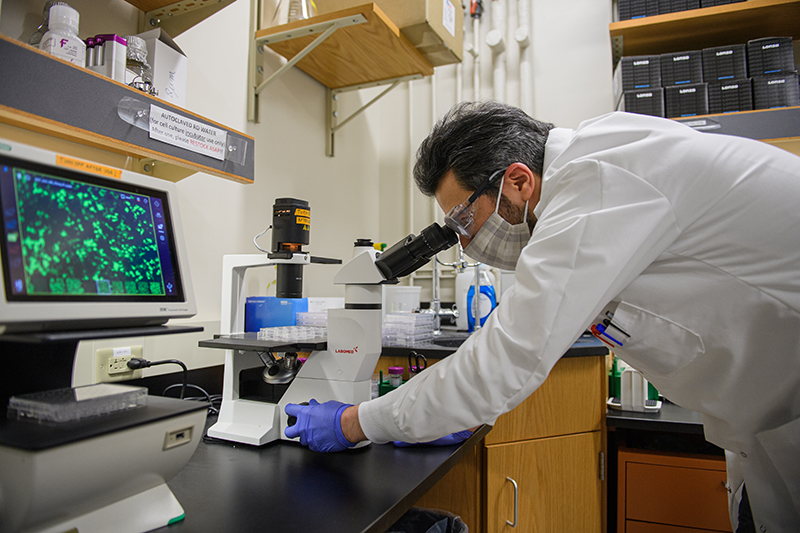
To identify possible drug targets, Majid Kazemian, assistant professor in the departments of computer science and biochemistry at Purdue University, said the research team examined more than 1,600 previously FDA-approved drugs with known targets.
We looked at the genes that are up-regulated by COVID-19 but down-regulated by specific drugs, and Ruxolitinib was the top drug with that property, he said.
Within the last few years, scientists have discovered that the immune complement system a complex system of small proteins produced by the liver that aids, or complements, the bodys antibodies in the fight against blood-borne pathogens can work inside cells and not just in the bloodstream.
Surprisingly, the study found that this response is triggered in cells of the small structures in the lungs known as alveoli, Kazemian said.
We observed that SARS-CoV2 infection of these lung cells causes expression of an activated complement system in an unprecedented way, Kazemian said. This was completely unexpected to us because we were not thinking about activation of this system inside the cells, or at least not lung cells. We typically think of the complement source as the liver.
Claudia Kemper, senior investigator and chief of the Complement and Inflammation Research Section of the National Institutes of Health, was among the first to characterize novel roles of the complement system in the immune system. She agreed these latest findings are surprising.
The complement system is traditionally considered a liver-derived and blood-circulating sentinel system that protects the host against infections by bacteria, fungi and viruses, she said. It is unexpected that in the setting of a SARS-CoV2 infection, this system rather turns against the host and contributes to the detrimental tissue inflammation observed in severe COVID-19. We need to think about modulation of this intracellular, local, complement when combating COVID-19.
Dr. Ben Afzali, an Earl Stadtman Investigator of the National Institute of Healths National Institute of Diabetes and Digestive and Kidney Diseases, said there are now indications that this has implications for difficulties in treating COVID-19.
These findings provide important evidence showing not only that complement-related genes are amongst the most significant pathways induced by SARS-CoV2 in infected cells, but also that activation of complement occurs inside of lung epithelial cells, i.e., locally where infection is present, he said.
This may explain why targeting the complement system outside of cells and in the circulation has, in general, been disappointing in COVID-19. We should probably consider using inhibitors of complement gene transcription or complement protein activation that are cell permeable and act intracellularly instead.
Afzali cautions that appropriate clinical trials should be conducted to establish whether a combination treatment provides a survival benefit.
The second finding that I think is important is that the data suggest potential benefit for patients with severe COVID-19 from combinatorial use of an antiviral agent together with an agent that broadly targets complement production or activation within infected cells, he said. These data are promising, but it is important to acknowledge that we carried out the drug treatment experiments in cell lines infected with SARS-CoV2. So, in and of themselves they should not be used to direct treatment of patients"
Kemper added that the unexpected findings bring more questions.
A currently unexplored and possibly therapeutically interesting aspect of our observations is also whether the virus utilizes local complement generation and activation to its benefit, for example, for the processes underlying cell infection and replication, she said.
About Purdue University
Purdue University is a top public research institution developing practical solutions to todays toughest challenges. Ranked the No. 5 Most Innovative University in the United States by U.S. News & World Report, Purdue delivers world-changing research and out-of-this-world discovery. Committed to hands-on and online, real-world learning, Purdue offers a transformative education to all. Committed to affordability and accessibility, Purdue has frozen tuition and most fees at 2012-13 levels, enabling more students than ever to graduate debt-free. See how Purdue never stops in the persistent pursuit of the next giant leap at https://purdue.edu/.
***
ABSTRACT
SARS-CoV2 drives JAK1/2-dependent local complement hyperactivation
Bingyu Yan1, Tilo Freiwald2,3,4, Daniel Chauss2, Luopin Wang5, Erin West3, Carmen Mirabelli6, Charles J Zhang7, Eva-Maria Nichols8, Nazish Malik8, Richard Gregory8, Marcus Bantscheff8, Sonja Ghidelli-Disse8, Martin Kolev8, Tristan Frum9, Jason R Spence9,10, Jonathan Z. Sexton7,9, Konstantinos D. Alysandratos11,12, Darrell N. Kotton11,12, Stefania Pittaluga13, Jack Bibby3, Nathalie Niyonzima14, Matthew R Olson15, Shahram Kordasti16,17, Didier Portilla2,18, Christiane E Wobus6, Arian Laurence19, Michail S Lionakis20, Claudia Kemper3,21*, Behdad Afzali2*, Majid Kazemian1,5*
- Department of Biochemistry, Purdue University, West Lafayette, IN, USA;
- Immunoregulation Section, Kidney Diseases Branch, National Institute of Diabetes and Digestive and Kidney Diseases (NIDDK), NIH, Bethesda, MD, USA;
- Complement and Inflammation Research Section (CIRS), National Heart, Lung, and Blood Institute (NHLBI), National Institutes of Health (NIH), Bethesda, MD, USA;
- Department of Nephrology, University Hospital Frankfurt, Goethe-University, Frankfurt, Germany;
- Department of Computer Science, Purdue University, West Lafayette, IN, USA;
- Department of Microbiology and Immunology, University of Michigan, Ann Arbor, MI, USA;
- Department of Medicinal Chemistry, College of Pharmacy, University of Michigan, Ann Arbor, MI, USA;
- GlaxoSmithKline, Stevenage, UK;
- Department of Internal Medicine, Gastroenterology, Michigan Medicine at the University of Michigan, Ann Arbor, MI, USA
- Department of Cell and Developmental Biology, University of Michigan, Ann Arbor, MI, USA;
- Center for Regenerative Medicine of Boston University and Boston Medical Center, Boston, MA, 1702118, USA;
- The Pulmonary Center and Department of Medicine, Boston University School of Medicine, Boston, MA, 02118, USA;
- Laboratory of Pathology, Center for Cancer Research, National Cancer Institute (NCI), NIH, Bethesda, MD, USA;
- Center of Molecular Inflammation Research (CEMIR), Department of Cancer Research and Molecular Medicine, Norwegian University of Science and Technology (NTNU), 7491 Trondheim, Norway;
- Department of Biological Sciences, Purdue University, West Lafayette, IN, USA;
- CRUK-KHP Centre, Comprehensive Cancer Centre, Kings College London, London, UK;
- Haematology Department, Guys Hospital, London, UK;
- Division of Nephrology and the Center for Immunity, Inflammation and Regenerative Medicine, University of Virginia, VA, USA
- Nuffield Department of Medicine, University of Oxford, UK;
- Fungal Pathogenesis Section, Laboratory of Clinical Immunology and Microbiology, National Institute of Allergy and Infectious Diseases (NIAID), NIH, Bethesda, MD, USA;
- Institute for Systemic Inflammation Research, University of Lübeck, Lübeck, Germany;
Patients with coronavirus disease 2019 (COVID-19) present a wide range of acute clinical manifestations affecting the lungs, liver, kidneys and gut. Angiotensin converting enzyme (ACE) 2, the best-characterized entry receptor for the disease-causing virus SARS-CoV2, is highly expressed in the aforementioned tissues. However, the pathways that underlie the disease are still poorly understood. Here, we unexpectedly found that the complement system was one of the intracellular pathways most highly induced by SARS-CoV2 infection in lung epithelial cells. Infection of respiratory epithelial cells with SARS-CoV2 generated activated complement component C3a and could be blocked by a cell-permeable inhibitor of complement factor B (CFBi), indicating the presence of an inducible cell-intrinsic C3 convertase in respiratory epithelial cells. Within cells of the bronchoalveolar lavage of patients, distinct signatures of complement activation in myeloid, lymphoid and epithelial cells tracked with disease severity. Genes induced by SARS-CoV2 and the drugs that could normalize these genes both implicated the interferon-JAK1/2-STAT1 signaling system and NF-kB as the main drivers of their expression. Ruxolitinib, a JAK1/2 inhibitor and the top predicted pharmaceutical candidate, normalized interferon signature genes and all complement gene transcripts induced by SARS-CoV2 in lung epithelial cell lines, but did not affect NF-kB-regulated genes. Ruxolitinib, alone or in combination with the anti-viral Remdesivir, inhibited C3a protein produced by infected cells. Together, we postulate that combination therapy with JAK inhibitors and drugs that normalize NF-kB-signaling could potentially have clinical application for severe COVID-19.
( Press Release Image: https://photos.webwire.com/prmedia/5/272525/272525-1.jpg )
WebWireID272525
This news content was configured by WebWire editorial staff. Linking is permitted.
News Release Distribution and Press Release Distribution Services Provided by WebWire.