Philips launches innovative 3D navigation system to enhance minimally invasive treatment of vascular disease
VesselNavigator has been designed for use in conjunction with Philips interventional X-ray systems to guide catheters during treatment of vascular disease.
Major reduction of contrast medium (70%) demonstrated in clinical study, enabling minimally invasive treatment of aortic aneurysms in the fast growing number of patients currently unable to benefit from minimally-invasive techniques.
Royal Philips (NYSE: PHG, AEX: PHIA) today announced the launch of VesselNavigator*, its latest innovation in live 3D catheter navigation to guide the minimally invasive treatment of patients with vascular diseases such as aortic aneurysms (ballooning of the aorta). This new catheter navigation solution, designed for use in conjunction with Philips interventional X-ray systems, enhances the precision and accuracy of stent placement, while at the same time significantly reducing contrast medium usage. As a result, minimally invasive treatment options will be available to patients previously unable to benefit from new image-guided intervention techniques.
Developed in collaboration with clinical partners such as the University Hospital Cologne (Germany) and the University Hospital Ghent (Belgium), VesselNavigator complements Philips current image-guided therapy portfolio within the field of endovascular and hybrid suite solutions. It addresses the need for advanced 3D live-image guidance solutions, as the treatment for vascular disease is experiencing a major transition from open surgery to minimally invasive procedures, with such procedure volumes growing at high single-digit rates.
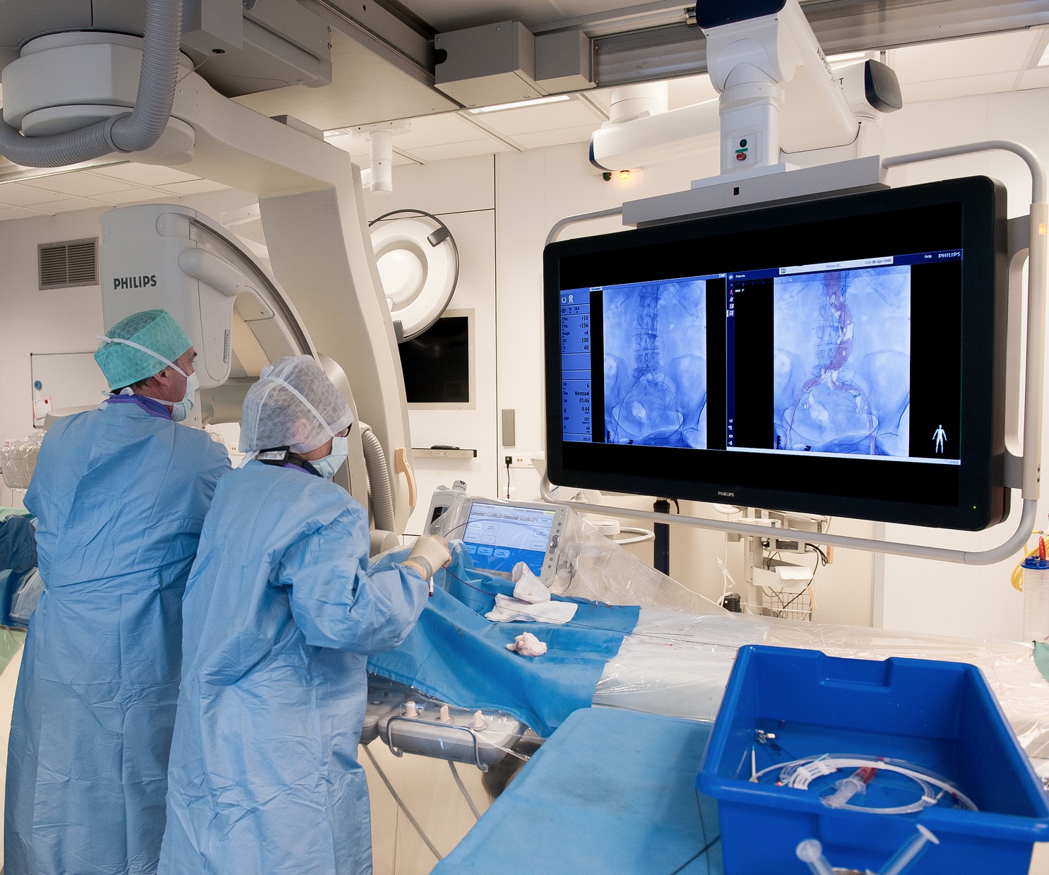
During endovascular procedures a catheter is maneuvered, with the aid of image guidance, through major arteries or veins in order to locally position and deploy implants such as stents to reinforce the wall of the affected blood vessel. Using conventional 2D X-ray image guidance, clinicians often perceive the visualization of the vessel anatomy during these procedures as if a dimension is missing, adding to procedure complexity. Many are more familiar with open surgery, during which they can physically see and touch the blood vessels they are trying to repair. VesselNavigator brings back the 3D anatomy they were used to seeing in open surgery.
VesselNavigator can be used for all types of endovascular procedures, but one of its key applications is guidance during the treatment of aortic aneurysms, which if left untreated could lead to severe complications such as massive internal bleeding. At the location of the aneurysm, the aorta often has smaller side branches, such as those that supply blood to the patients kidneys. Custom-made stents are therefore often made with dedicated openings that need to be precisely registered with these feeding vessels in order to repair the aorta and maintain critical blood flow to other abdominal organs. With conventional X-ray imaging it is very challenging to position the stent in the precise orientation. Endovascular aortic aneurysm repair is therefore a very complex procedure, and the more time it takes, the more contrast medium is needed for X-ray visualization and guidance in order to succeed.
VesselNavigator fuses live interventional X-ray images with pre-acquired 3D MRI or CT images of the patients vascular structures. The resulting 3D color-coded images of the vessels provide enhanced real-time visual guidance, making it easier to maneuver through the vascular network without the need to enhance the X-ray visualization with the repeated use of an injected contrast medium. In recent studies, VesselNavigator has been shown to reduce contrast medium usage by 70%[1] and procedure times by 18%[2], contributing to more patient friendly, more efficient and more cost effective treatment of vascular conditions.
VesselNavigator gives vascular surgeons during endovascular procedures the 3D view of the patients anatomy, which they are familiar with from open surgery. It also significantly reduces the amount of contrast medium required, which means a lesser burden on the kidneys. And with a growing population of elderly and diabetic people who suffer from poor kidney function, reducing contrast medium requirements will open up endovascular treatments to a wider range of patients, says Professor Dr. Frank Vermassen, Head of Vascular and Thoracic Surgery at University Hospital Ghent.
The strong growth in image-guided therapy procedures is driven by the significant benefits they offer for healthcare systems and patients, including reduced patient trauma, shorter hospital stays, and lower health care costs, said Bert van Meurs, General Manager Image Guided Therapy at Philips. It is an area where technology innovation and clinical innovation go hand in hand. VesselNavigator shows our commitment to in close collaboration with leading clinical and industrial partners advance the development of innovative technologies that enable less invasive, more accurate and localized therapies.
VesselNavigator joins Philips extensive portfolio of live, 3D image-guided navigation solutions for image-guided minimally invasive therapies. This portfolio also includes HeartNavigator and EchoNavigator for structural heart disease repairs, EP Navigator for cardiac electrophysiology interventions, and EmboGuide to support tumor embolization in cancer treatment. Philips leading position in image-guided therapies was recently further enhanced by the acquisition of Volcano Corporation, a global leader in catheter-based imaging and measurement solutions for cardiovascular applications.
* Not available for sale in the U.S. pending 510(k) clearance
[1] Ref Pubmed: Tacher et al; J Vasc Interv Radiol. 2013 Nov;24(11):1698-706
[2] Ref Pubmed: Sailer et al; Eur J Vasc Endovasc Surg. 2014 Apr;47(4):349-56
( Press Release Image: https://photos.webwire.com/prmedia/2/197415/197415-1.jpg )
WebWireID197415
This news content was configured by WebWire editorial staff. Linking is permitted.
News Release Distribution and Press Release Distribution Services Provided by WebWire.